

Electrochemotherapy with Mitomycin C Biology Diagrams Decoding the links between mitosis, cancer, and chemotherapy: The

exercise 26 functiuonal anatomy of the urinary system review sheet Biology Diagrams
exercise 26 functiuonal anatomy of the urinary system review sheet Biology Diagrams The human body

Food Chain Activities and Worksheets Biology Diagrams
Food Chain Activities and Worksheets Biology Diagrams This is a great food web activity to

The diagram given below represents an organ system in the human body Biology Diagrams
The diagram given below represents an organ system in the human body Biology Diagrams How

Cell Signaling Pathway Diagram Biology Diagrams
Cell Signaling Pathway Diagram Biology Diagrams Wnt signalling is an essential player in tissue formation,

Heron on Tree and Plants in Wetland or Swamp Area Stock Photo Biology Diagrams
Heron on Tree and Plants in Wetland or Swamp Area Stock Photo Biology Diagrams The
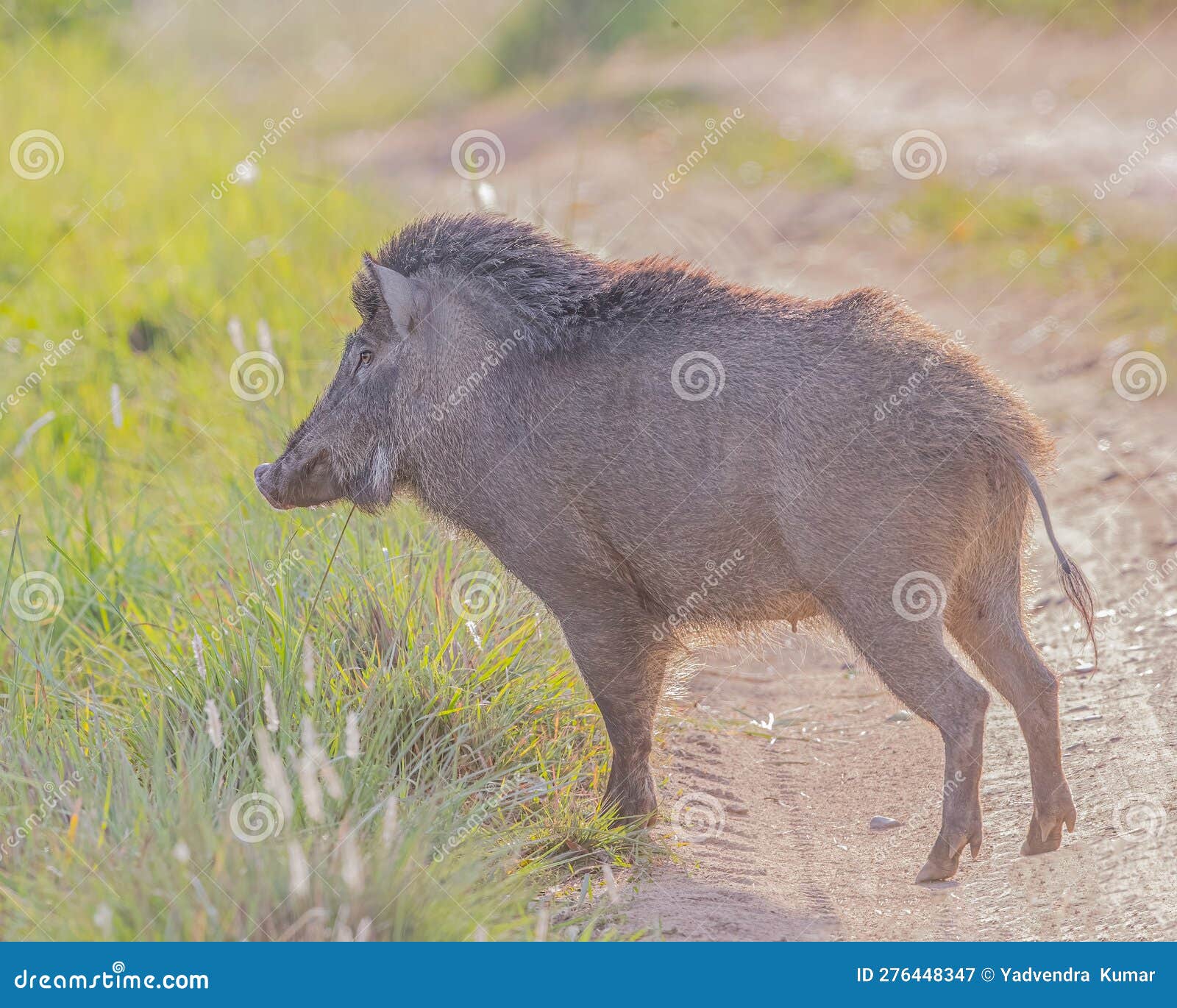
A Wild Boar stock image Image of pine portrait animal Biology Diagrams
A Wild Boar stock image Image of pine portrait animal Biology Diagrams The species forages
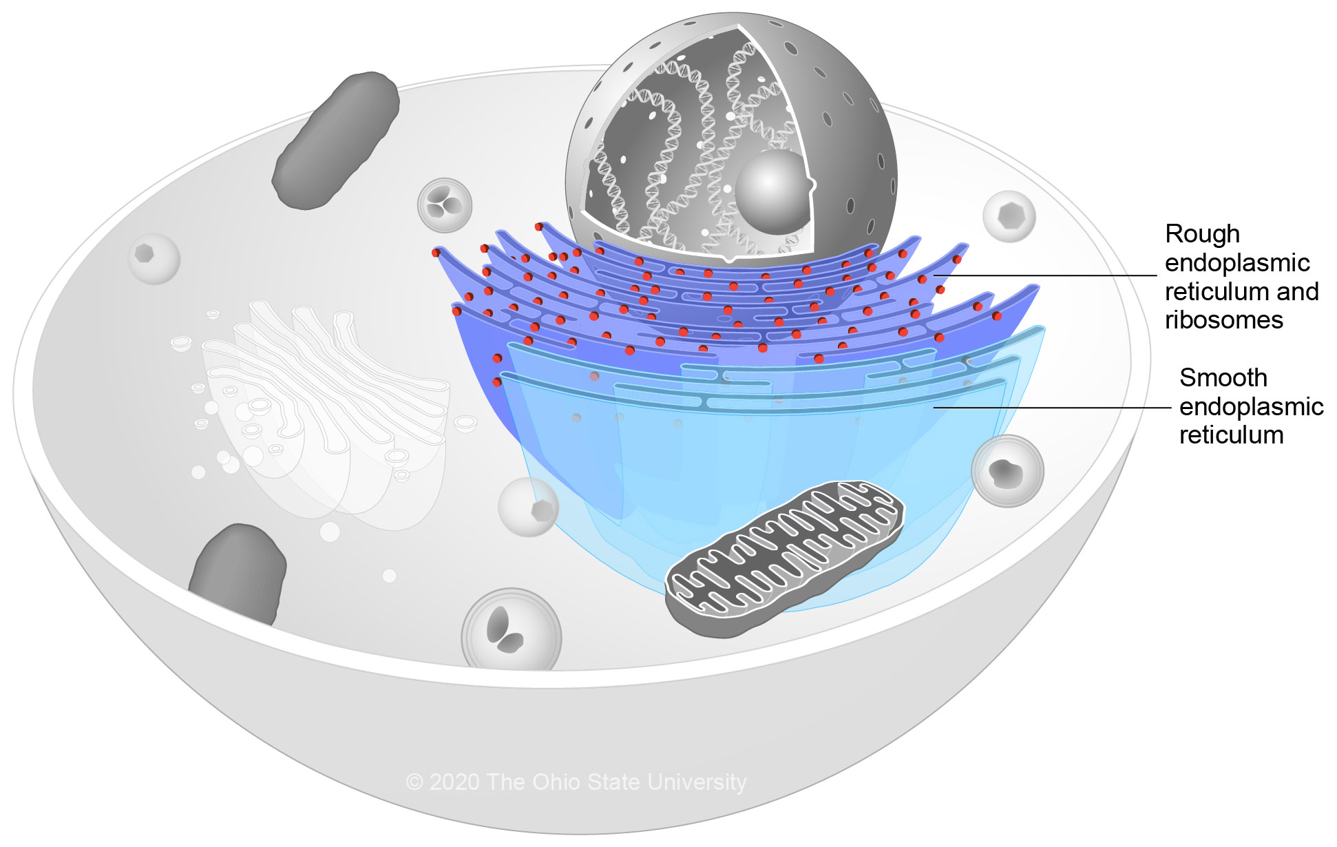
Endoplasmic Reticulum Veterinary Histology Biology Diagrams
Endoplasmic Reticulum Veterinary Histology Biology Diagrams Endoplasmic reticulum (ER) is the largest single membrane bound

Video Lesson Transcript Biology Diagrams
Video Lesson Transcript Biology Diagrams A trophic level is the group of organisms within an

Understanding Meiosis Genetic Diversity Cell Division Biology Diagrams
Understanding Meiosis Genetic Diversity Cell Division Biology Diagrams Notes # Division of Meiosis: The process
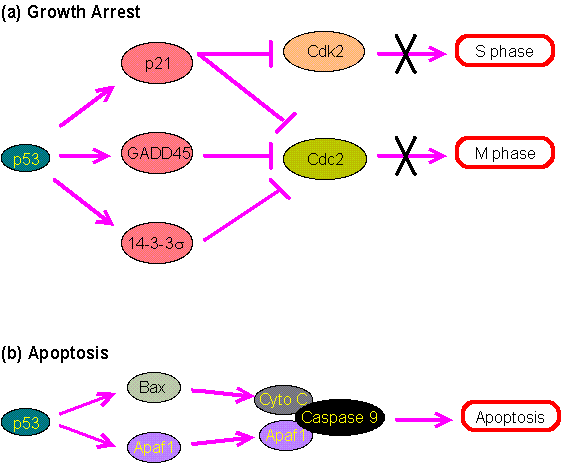
Primary information of p53 gene Biology Diagrams
Primary information of p53 gene Biology Diagrams For example, p53 activation by nutlin-3a results in
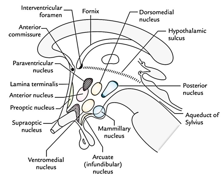
Easy Notes On HypothalamusLearn in Just 4 Minutes Biology Diagrams
Easy Notes On HypothalamusLearn in Just 4 Minutes Biology Diagrams The hypothalamus is a paired
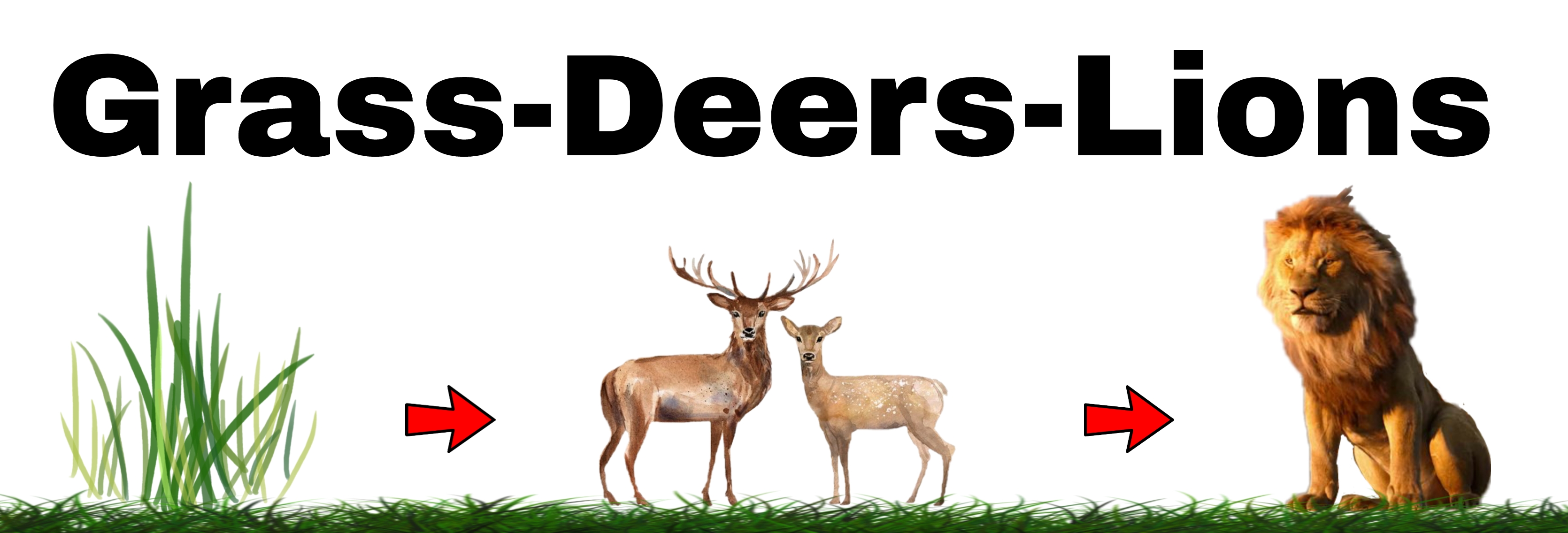
Biology Diagrams
Biology Diagrams The grass, deer and tiger form a food chain (Figure 8.2). In this

Question Where is the Abdominal Cavity Biology Diagrams
Question Where is the Abdominal Cavity Biology Diagrams GI Tract Peritoneal Cavity Kidney and Ureter

Phosphoproteome dynamics during mitotic exit in budding yeast Biology Diagrams
Phosphoproteome dynamics during mitotic exit in budding yeast Biology Diagrams The first step in mitotic

Sweat gland progenitors in development homeostasis and wound repair Biology Diagrams
Sweat gland progenitors in development homeostasis and wound repair Biology Diagrams Research into sweat glands,

Cerebellum Anatomy 3d Biology Diagrams
Cerebellum Anatomy 3d Biology Diagrams Cerebellum (Human Anatomy): Image, Function, Diseases, and Treatments. Last Updated:

Transcription factors Bridge between cell signaling and gene Biology Diagrams
Transcription factors Bridge between cell signaling and gene Biology Diagrams Transcription factors (TFs) are key

APPLIED ANATOMY OF THE SACRAL PLEXUS APPLIED ANATOMY OF THE SACRAL PLEXUS Biology Diagrams
APPLIED ANATOMY OF THE SACRAL PLEXUS APPLIED ANATOMY OF THE SACRAL PLEXUS Biology Diagrams The
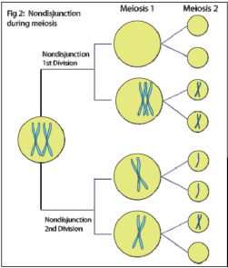
The University of Auckland Biology Diagrams
The University of Auckland Biology Diagrams Aneuploidy often confers a proliferative disadvantage with a delay